Life History of Western Tussock Moths, Orgyia vetusta
with Parasitoid Activity
Joyce Gross
University of California at Berkeley
Occasional Papers of the Moth Photographers Group
Number 2 - 30 January 2007
mothphotographersgroup.msstate.edu/Files/Live/JGross/TussockMothParasitoids.htm
|

|
|
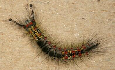
One of the Tussock Moth caterpillars from the side of Valley Life Sciences Building (VLSB). Thanks to Peter Ralph for finding this caterpillar, and for noticing others were pupating. Dozens if not hundreds of these caterpillars have been busy eating leaves on a Coast Live Oak next to the building where this caterpillar was found. Photos taken May 10, 2006.
Taxonomy: These moths have been known as Orgyia vetusta, but according to Jerry Powell "there is some disagreement, and that name maybe should be limited to the immediate coastal populations, in which case ours is O. cana. The larvae differ."
|
|

|

|
This is a caterpillar making its cocoon on the side of VLSB. Photo taken June 15, 2006. Beside it is a completed cocoon (taken from the side of VLSB; I brought several home). This is what you would see attached to the wall or a tree. The caterpillar not only spins around and above it a network of silk but plucks hairs from its own body to incorporate into the completed covering. It then enters the pupal stage of the life cycle.
|
|

|

|

|
In the leftmost photo, the dark junk on the left side is a bit of the exoskeleton, particularly the caterpillar's head/mouth parts. The center photo shows another pupa with its cocoon. (It was hard to keep them inside the cocoons after removing from the wall.) The former caterpillar mouthparts-head-exoskeleton are especially noticeable towards the left bottom in this photo. Note also the former caterpillar tufts still visible on the pupa. The caterpillar whose pupal remains are shown in the rightmost photo was inoculated with the egg of a parasitoid fly. The pupa is dead, a hollow shell, and the pupa of the parasitoid larva that developed internally in the moth pupa is on the right. Photos taken June 6, 2006.
|
|

|

|
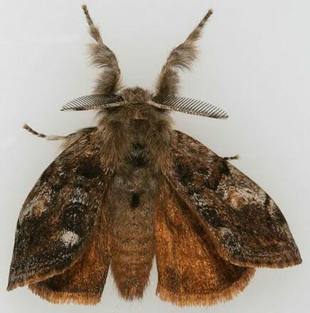
Adult females are wingless. When they emerge from the cocoon (eclosure) they remain upon it and release a pheromone, a sex-attractant that brings males to her. The large antenna of the male is very sensitive to the species-specific pheromone and any males downwind of the female will quickly follow the scent to her location. Photos take June 12, 2006.
|
|

|

|
The first two adults that eclosed at my house are here shown mating. This female produced some eggs afterwards but not as many as the ones I saw still attached to VLSB. Photo taken June 14, 2006. At right is an undisturbed female finishing laying eggs attached to the side of VLSB and on her cocoon. She is on the left clinging to her cocoon and egg mass. The eggs are being covered with hair taken from the mother's body. The cocoon from which she emerged is towards the top. Photo taken June 15, 2006.
|
|

|

|
Some of last year's eggs, most of which hatched, remain on the wall of the VLSB. The old cocoon and pupal case are still visible. In the second photo can be seen a portion of an Earwig using an old cocoon for shelter. The white material is paint that had been applied to the building.
|
|

|

|
The fly parasitoid has been identified by Norm Woodley as Compsilura concinnata, a member of the Tachinidae.
.
|
Notes:
Joyce Gross is a programmer at The Biodiversity Sciences Technology group where she develops web-based systems and databases for biological and natural history museum collections, biodiversity science projects, and other research at the University of California, Berkeley.
References:
California Moth Specimens Database records for Lymantriidae.
Ferguson, Douglas C. 1978. The Moths of America North of Mexico, Fascicle 22.2, Noctuoidea (in part): Lymantriidae. E. W. Classey Ltd. and The Wedge Entomological Research Foundation. London. pp.110, 8 plates.
|